Nervous tissue structure, functions, drawing. Nervous tissue: functions, structure. Properties of nervous tissue. Cellular composition of nervous tissue
MAIN QUESTIONS OF THE TOPIC:
1. General morphofunctional characteristics of nervous tissue.
2. Embryonic histogenesis. Differentiation of neuroblasts and glioblasts. The concept of regeneration of structural components of nervous tissue.
3. Neurocytes (neurons): sources of development, classification, structure, regeneration.
4. Neuroglia. General characteristics. Sources of gliocyte development. Classification. Macroglia (oligodendroglia, astroglia and ependymal glia). Microglia.
5. Nerve fibers: general characteristics, classification, structure and functions of unmyelinated and myelinated nerve fibers, degeneration and regeneration of nerve fibers.
6. Synapses: classifications, structure of the chemical synapse, structure and mechanisms of excitation transmission.
7. Reflex arcs, their sensitive, motor and associative links.
BASIC THEORETICAL PROVISIONS
NERVOUS TISSUE
Nervous tissue performs the functions of perception, conduction and transmission of excitation received from external environment and internal organs, as well as analysis, storage of received information, integration of organs and systems, interaction of the body with the external environment.
Basic structural elements nervous tissue - cells And neuroglia.
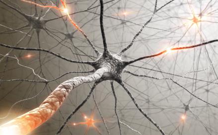
Neurons
Neurons consist of a body ( perikarya) and processes, among which are dendrites And axon(neuritis). There can be many dendrites, but there is always one axon.
A neuron, like any cell, consists of 3 components: nucleus, cytoplasm and cytolemma. The main volume of the cell is in the processes.
 Core
occupies a central position in perikaryone. One or several nucleoli are well developed in the nucleus.
Core
occupies a central position in perikaryone. One or several nucleoli are well developed in the nucleus.
Plasmolemma takes part in the reception, generation and conduction of nerve impulses.
Cytoplasm the neuron has a different structure in the perikaryon and in the processes.
The cytoplasm of the perikaryon contains well-developed organelles: ER, Golgi complex, mitochondria, lysosomes. Neuron-specific cytoplasmic structures at the light-optical level are chromatophilic substance of cytoplasm and neurofibrils.
 Chromatophilic substance cytoplasm (Nissl substance, tigroid, basophilic substance) appears when nerve cells are stained with basic dyes (methylene blue, toluidine blue, hematoxylin, etc.) in the form of granularity - these are accumulations of grEPS cisterns. These organelles are absent in the axon and axon hillock, but are present in the initial segments of dendrites. The process of destruction or disintegration of clumps of basophilic substance is called tigrolysis and is observed during reactive changes in neurons (for example, when they are damaged) or during their degeneration.
 Neurofibrils is a cytoskeleton consisting of neurofilaments and neurotubules that form the framework of the nerve cell. Neurofilaments represent intermediate filaments 8-10 nm in diameter, formed by fibrillar proteins. The main function of these cytoskeletal elements is support - to ensure a stable shape of the neuron. A similar role is played by subtle microfilaments(transverse diameter 6-8 nm), containing actin proteins. Unlike microfilaments in other tissues and cells, they do not bind to micromyosins, which makes active contractile functions in mature nerve cells impossible.
Neurotubules according to the basic principles of their structure, they are actually no different from microtubules. They, like all microtubules, have a transverse diameter of about 24 nm; the rings are closed by 13 molecules of the globular protein tubulin. In nervous tissue, microtubules perform a very important, if not unique role. As elsewhere, they have a frame (support) function and provide cyclosis processes. Microtubes are polar. It is the polarity of the microtube, which has negatively and positively charged ends, that makes it possible to control diffusion-transport flows in the axon (the so-called fast and slow axotok). Their detailed description we will give below.
In addition, lipid inclusions (lipofuscin grains) can be seen quite often in neurons. They are characteristic of old age and often appear during degenerative processes. Some neurons normally exhibit pigment inclusions (for example, with melanin), which causes staining of the nerve centers containing similar cells (substantia nigra, bluish spot).
Neurons are energetically extremely dependent on aerobic phosphorylation and in adulthood are virtually incapable of anaerobic glycolysis. In this regard, nerve cells are strongly dependent on the supply of oxygen and glucose, and if blood flow is disrupted, nerve cells almost immediately cease their vital functions. The moment blood flow in the brain stops means the beginning of clinical death. With instant death, at room temperature, and normal body temperature, the processes of self-destruction in neurons are reversible within 5-7 minutes. This is the period of clinical death, when the body can be revived. Irreversible changes in nervous tissue lead to the transition from clinical death to biological death.
In the body of neurons one can also see transport vesicles, some of which contain mediators and modulators. They are surrounded by a membrane. Their size and structure depend on the content of a particular substance.
Dendrites- short shoots, often highly branched. Dendrites in the initial segments contain organelles similar to the body of a neuron. The cytoskeleton is well developed.
Axon(neurite) is most often long, weakly branched or not branched. It lacks grEPS. Microtubules and microfilaments are arranged in an orderly manner. Mitochondria and transport vesicles are visible in the cytoplasm of the axon. The axons are mostly myelinated and surrounded by processes of oligodendrocytes in the central nervous system, or lemmocytes in the peripheral nervous system. The initial segment of the axon is often expanded and is called the axon hillock, where the summation of signals entering the nerve cell occurs, and if the exciting signals are of sufficient intensity, then an action potential is formed in the axon and the excitation is directed along the axon, transmitted to other cells (action potential).
Axotok (axoplasmic transport of substances). Nerve fibers have a unique structural apparatus - microtubules, through which substances move from the cell body to the periphery ( anterograde axotoc) and from the periphery to the center ( retrograde axotoc).
There are fast (at a speed of 100-1000 mm/day) and slow (at a speed of 1-10 mm/day) axotoc. Fast Aksotok– the same for different fibers; requires a significant concentration of ATP; occurs with the participation of transport vesicles. It transports mediators and modulators. Slow Aksotok– due to it, biologically active substances, as well as components of cell membranes and proteins, spread from the center to the periphery.
Nerve impulse transmitted along the neuron membrane in a certain sequence: dendrite – perikaryon – axon.
Classification of neurons
 1. According to morphology (by the number of processes) there are:
- multipolar neurons (d) - with many processes (the majority of them in humans),
- unipolar neurons (a) - with one axon,
- bipolar neurons (b) - with one axon and one dendrite (retina, spiral ganglion).
- false- (pseudo-) unipolar neurons (c) - dendrite and axon extend from the neuron as one process, and then separate (in the dorsal ganglion). This is a variant of bipolar neurons.
2. By function (by location in reflex arc) are distinguished:
 - afferent (sensitive) neurons (arrow on the left) - perceive information and transmit it to the nerve centers. Typical sensitive ones are pseudounipolar and bipolar neurons of the spinal and cranial ganglia;
- associative (insert) neurons interact between neurons, most of them are in the central nervous system;
- efferent (motor) neurons (arrow on the right) generate a nerve impulse and transmit excitation to other neurons or cells of other types of tissue: muscle, secretory cells.
Synapses
Synapses - these are specific neuron contacts that ensure the transfer of excitation from one nerve cell to another. Depending on the methods of transmission of excitation, chemical and electrical synapses are distinguished.
Evolutionarily more ancient and primitive are electrical synaptic contacts . Their structure is close to gap-like contacts (nexuses). It is believed that the exchange occurs in both directions, but there are cases when excitation is transmitted in one direction. Such contacts are often found in lower invertebrates and chordates. In mammals, electrical contacts have great importance in the process of interneuron interactions in the embryonic period of development. This type of contact in adult mammals occurs in limited areas, for example they can be seen in the mesencephalic nucleus of the trigeminal nerve.
Chemical synapses . Chemical synapses use special substances to transmit excitation from one nerve cell to another - mediators, from which they got their name. In addition to mediators, they also use modulators. Modulators are special chemical substances, which themselves do not cause excitation, but can either enhance or weaken sensitivity to mediators (that is, modulate the threshold sensitivity of the cell to excitation).
Chemical synapse provides unidirectional transfer of excitation. Structure of a chemical synapse:
1) Presynaptic zone– presynaptic extension, most often representing the axon terminal, which contains synaptic vesicles, cytoskeletal elements (neurotubules and neurofilaments), mitochondria;
2) Synaptic cleft, which receives mediators from the presynaptic zone;
3) Postsynaptic zone is an electron-dense substance with receptors for a transmitter on the membrane of another neuron .

FILM SYNAPSES
Classification of synapses
:
1. Depending on what structures of two neurons interact at the synapse, we can distinguish:
Axo-dendritic (presynaptic structure - axon, postsynaptic - dendrite);
Axo-axonal;
Axo-somatic.
2. By function they distinguish:
- stimulating synapses that lead to depolarization of the postsynaptic membrane and activation of the nerve cell;
- inhibitory synapses, which lead to hyperpolarization of the membrane, which reduces the threshold sensitivity of the neuron to external influences.
3. Based on the main transmitter contained in synaptic vesicles, synapses are divided into groups:
- Cholinergic (acetylcholinergic): excitatory and inhibitory;
- Adrenergic (monoaminergic, noradrenergic, dopaminergic): mainly excitatory, but also inhibitory;
- Serotonergic (sometimes attributed to the previous group): excitatory;
- GABAergic (mediator gamma-aminobutyric acid): inhibitory;
- Peptidergic (mediators - a large group of substances, mainly: vasointerstitial polypeptide, vasopressin, substance P (pain mediator), neuropeptide Y, oxytocin, beta-endorphin and enkephalins (anti-pain), dynorphin, etc.).
Synaptic vesicles separated from the hyaloplasm by a single membrane. Choline-containing vesicles are electron-clear, 40-60 µm in diameter. Adrenaceous - with an electron-dense core, a light rim, with a diameter of 50-80 microns. Glycine-containing and GABA-containing - have an oval shape. Peptide-containing - with an electron-dense core, a light border, with a diameter of 90-120 microns.
The mechanism of excitation transmission in a chemical synapse: an impulse arriving along an afferent fiber causes excitation in the presynaptic zone and leads to the release of a transmitter through the presynaptic membrane. The transmitter enters the synaptic cleft. On the postsynaptic membrane there are receptors for the neurotransmitter (cholinergic receptors for the mediator acetylcholine; adrenergic receptors for norepinephrine). Subsequently, the connection between the mediators and the receptors is broken. The mediator is either metabolized, or undergoes reabsorption by presynaptic membranes, or is captured by the membranes of astrocytes with subsequent transfer of the mediator to nerve cells.
Neuronal regeneration. Neurons are characterized only by intracellular regeneration. They are a stable population of cells and do not divide under normal conditions. But there are exceptions. Thus, the ability to divide has been proven in nerve cells in the epithelium of the olfactory analyzer, in some ganglia (clusters of neurons of the autonomic nervous system) animals.
Neuroglia
Neuroglia
- a group of cells of nervous tissue located between neurons, distinguished microglia and macroglia
.
Macroglia
Macroglia CNS is divided into the following cells: astrocytes (fibrous and protoplasmic), oligodendrocytes and ependymocytes (including tanycytes).
Macroglia of the peripheral nervous system: satellite cells and lemmocytes (Schwann cells).
Functions of macroglia: protective, trophic, secretory.
Astrocytes – stellate cells, numerous processes of which branch and surround other brain structures. Astrocytes are found only in the central nervous system and analyzers - derivatives of the neural tube.
Types of astrocytes: fibrous and protoplasmic astrocytes.
The process terminals of both cell types have buttonlike extensions (astrocytic feet), most of which terminate in the perivascular space, surrounding the capillaries and forming perivascular glial membranes.
Fibrous astrocytes have numerous, long, thin, weakly or not at all branched processes. Mainly present in the white matter of the brain.
Protoplasmic astrocytes are distinguished by short, thick and highly branched processes. Found predominantly in the gray matter of the brain. Astrocytes are located between the bodies of neurons, the unmyelinated and myelinated parts of the nerve processes, synapses, blood vessels, and subependymal spaces, isolating and at the same time structurally connecting them.
A specific marker of astrocytes is glial fibrillary acidic protein, from which intermediate filaments are formed.
Astrocytes have relatively large, light-colored nuclei, with a poorly developed nucleolar apparatus. The cytoplasm is weakly oxyphilic, aEPS and grEPS, and the Golgi complex are poorly developed in it. There are few mitochondria, they are small in size. The cytoskeleton is moderately developed in protoplasmic and well developed in fibrous astrocytes. There are a significant number of gap-like and desmosome-like contacts between cells.
In the postnatal period of human life, astrocytes are capable of migration, especially to areas of damage, and are capable of proliferation (from which benign astrocytoma tumors are formed).
Main functions of astrocytes: participation in blood-brain and cerebrospinal fluid barriers(with their processes they cover capillaries, the surfaces of the brain and participate in the transport of substances from vessels to neurons and vice versa), in connection with this they perform protective, trophic, regulatory functions; phagocytosis of dead neurons, secretion of biologically active substances: FGF, angiogenic factors, EGF, interleukin-I, prostaglandins.
Oligodendrocytes – cells with a small number of processes , capable of forming myelin sheaths around the bodies and processes of neurons. Oligodendrocytes are found in the gray and white matter of the central nervous system; in the peripheral nervous system there are types of oligodendrocytes - lemmocytes (Schwann cells). Oligodendrocytes and their varieties are characterized by the ability to form membrane duplications - mesaxon, which surrounds the process of a neuron, forming a myelin or non-myelin sheath.
The nuclei of oligodendrocytes are small, round, dark-colored, the processes are thin, do not branch or weakly branch. At the electron-optical level, organelles are well developed in the cytoplasm, especially the synthetic apparatus; the cytoskeleton is poorly developed.
Some oligodendrocytes are concentrated in close proximity to the nerve cell bodies ( satellite or mantle oligodendrocytes). The terminal zone of each process participates in the formation of a nerve fiber segment, that is, each oligodendrocyte provides an environment for several nerve fibers at once.
Lemmocytes (Schwann cells ) peripheral nervous system are characterized by elongated, dark-colored nuclei, poorly developed mitochondria and synthetic apparatus (granular, smooth ER, lamellar complex). Lemmocytes surround the processes of neurons in the peripheral nervous system, forming myelin or non-myelin sheaths. In the area of formation of the roots of the spinal and cranial nerves, lemmocytes form clusters (glial plugs), preventing the penetration of the processes of associative neurons of the central nervous system beyond its boundaries.
In the peripheral nervous system, in addition to lemmocytes, There are other types of oligodendrocytes: satellite (mantle) gliocytes in the peripheral ganglia around the cell bodies of neurons, gliocytes of nerve endings, specific morphological features which are considered in the study of nerve endings and the anatomy of nerve ganglia.
The main functions of oligodendrocytes and their varieties: forming myelin or non-myelin sheaths around neurons, providing insulating, trophic, supporting, protective functions; participate in the conduction of nerve impulses, in the regeneration of damaged nerve cells, phagocytosis of the remains of axial cylinders and myelin when the axon structure is disturbed distal to the site of damage.
Ependymocytes , or ependymal glia - low-prismatic cells that form a continuous layer covering the cavities of the brain. Ependymocytes are closely adjacent to each other, forming tight, slit-like and desmosomal contacts. The apical surface contains cilia, which in most cells are then replaced by microvilli. The basal surface has basal invaginations (invaginations), as well as long thin processes (from one to several), which penetrate to the perivascular spaces of the brain microvessels.
The cytoplasm of ependymocytes contains mitochondria, a moderately developed synthetic apparatus, a well-represented cytoskeleton, and a significant number of trophic and secretory inclusions.
Variants of ependymal glia are tanycytes . They line the choroid plexuses of the ventricles of the brain, the subcommissural organ of the posterior commissure. Actively participate in the formation of cerebrospinal fluid (CSF). They are characterized by the fact that the basal part contains thin long processes.
Main functions of ependymocytes: secretory (synthesis of cerebrospinal fluid), protective (providing blood-cerebrospinal fluid barrier), supporting, regulatory (tanycyte precursors direct the migration of neuroblasts in the neural tube in the embryonic period of development).
Microglia
Microgliocytes, or neural macrophages – small cells of mesenchymal origin (monocyte derivatives), diffusely distributed in the central nervous system, with numerous highly branching processes, are capable of migration. Microgliocytes are specialized macrophages of the nervous system. Their nuclei are characterized by a predominance of heterochromatin. Many lysosomes and lipofuscin granules are found in the cytoplasm; the synthetic apparatus is moderately developed.
Functions of microglia: protective (including immune).
Nerve fibers
A nerve fiber consists of a neuron extension - axial cylinder(dendrite or axon) and oligodendrocyte membrane or its variants.
Types of nerve fibers:
1) Depending on how the sheath was formed, nerve fibers are divided into myelin And unmyelinated.
In the peripheral nervous system, nerve fibers surround lemmocytes. One lemmocyte is associated with one nerve fiber. In the central nervous system, neuronal processes are surrounded by oligodendrocytes. Each oligodendrocyte participates in the formation of several nerve fibers.
Myelination fibers are carried out by lengthening and “winding” the mesaxon around the nerve cell process (in the peripheral nervous system) or lengthening and rotation of the oligodendrocyte process around the axial cylinder in the central nervous system.
myelin (meat) fibers in the peripheral nervous system consist of one neuron process, surrounded by an elongated lemmocyte duplicature (mesaxon). In the myelin fiber, mesaxon wraps repeatedly around the axial cylinder, forming multiple turns of the membrane - myelin. Zones of myelin loosening (penetration of lemmocyte cytoplasm) are called notches(Schmidt-Lanterman). Each lemmocyte forms a fiber segment, the areas of the borders of neighboring cells are unmyelinated and are called Ranvier interceptions Thus, along the length of the fiber, the myelin sheath has an intermittent course. The myelin sheath is a biological insulator. The propagation of depolarization in the myelin fiber occurs in jumps from interception to interception.
Unmyelinated (mellless) fibers in the peripheral nervous system consist of one or more axial cylinders embedded in the cytolemma of the surrounding lemmocyte. Mesaxon (membrane duplication) is short. Transmission of excitation in unmyelinated fibers occurs along the surface of the nerve through a change in surface charge.
2) Depending on the speed of the nerve impulse, the following types of nerve fibers are distinguished:
- Type A has subgroups:
- Aa- have the highest speed of excitation - 70-120 m/s (somatic motor nerve fibers);
- Ab— the conduction speed is 40-70 m/s. These are somatic afferent nerves and some efferent somatic nerves;
- Ag- conduction speed is 15-40 m/s - afferent and efferent sympathetic and parasympathetic nerves;
- Ad(delta) - conduction speed 5-18 m/s. This group of afferent somatic nerves transmits primary (fast) pain.
- Type B – conduction speed from 3 to 14 m/s – preganglionic sympathetic fibers, some parasympathetic fibers, that is, these are autonomic nerves.
- Type C – conduction speed 0.5-3 m/s: postganglionic autonomic fibers (unmyelinated). Conduct pain impulses of slow secondary pain (from the receptors of the dental pulp).
Neurogenesis. On days 15-17 of human intrauterine development, under the inducing influence of the notochord from primary ectoderm a neural plate (an accumulation of longitudinally lying cellular material) is formed. From days 17 to 21, the plate invaginates and first turns into neural groove, and then in phone. By the 25th day of embryogenesis, the neural tube splits off from the ectoderm and the anterior and posterior openings (neuropores) close. On the sides of the neural groove are located neural crest structures.
In the early stages of development, the neural tube is formed meduloblasts – stem cells of the nervous tissue of the central nervous system. Formed from the neural crest ganglionic plate consisting of ganglioblasts– stem cells of neurons and neuroglia of the peripheral nervous system. Meduloblasts and ganglioblasts intensively immigrate, divide and then differentiate.
In the early stages of intrauterine development, the neural tube is a layer of process cells lying in the form of one layer, but in several rows. They are limited internally and externally by limiting membranes. On the inner surface (adjacent to the cavity of the neural tube), meduloblasts divide.
Subsequently the neural tube forms several layers . Among them are:
- Internal limiting membrane: separates the cavity of the neural tube from the cells;
- Ependymal layer(ventricular in the area of the brain vesicles) is represented by blast precursor cells of macroglia;
- Subventricular zone(only in the anterior medullary vesicles), where neuroblast proliferation occurs;
- Mantle (cloak) layer, containing migrating and differentiating neuroblasts and glioblasts;
- Marginal layer(marginal veil) is formed by processes of glioblasts and neuroblasts. In it you can see the bodies of individual cells.
- External limiting membrane.
Differentons of nervous tissue of the central nervous system
- Differenton of a neuron: meduloblast – neuroblast – young neuron – mature neuron.
- Astrocyte differential: meduloblast – spongioblast – astroblast – protoplasmic or fibrous astrocyte.
- Oligodendrocyte diferron: meduloblast - spongioblast - oligodendroblast - oligodendrocyte.
- Differenton of ependymal glia: meduloblast – ependymoblast – ependymocyte or tanycyte.
- Differenton of microglia: blood stem cell – semi-stem blood cell (CFU GEMM) – CFU GM – CFU M – monoblast – promonocyte – monocyte – resting microgliocyte – activated microgliocyte.
Differentons of nervous tissue in the peripheral nervous system
1. Differenton of a neuron: ganglioblast – neuroblast – young neuron – mature neuron.
2. Lemmocyte differential: ganglioblast – glioblast – lemmocyte (Schwann cell).
Mechanisms of neurogenesis. During intrauterine development, neuroblasts migrate to the anatomical areas of the nerve centers. At the same time, they stop sharing. In the CNS, the migration of neuroblasts is controlled by adhesive intercellular interactions (with the help of cadherins and integrins of radial glia), signaling molecules of the intercellular substance (including fibronectins and laminins). After neuroblasts reach their permanent localization area, they begin to differentiate and form processes. The direction of growth of processes is also controlled by the mentioned adhesion molecules (cadherins, integrins, signaling molecules of the intercellular substance).
During intrauterine development and after birth, competitive interaction occurs between similar neurons of the nerve centers. In this case, nerve cells that do not have time to occupy the corresponding zone or form contacts undergo apoptosis. In early development, from a third to a half of nerve cells die.
In subsequent development, a glial environment is formed around the nerve cells and myelination of nerve fibers occurs. Nerve cells continue to form processes and synaptic contacts until puberty. Nervous tissue reaches its maximum development by the age of 25-30.
With age, the death of some nerve cells and compensatory hypertrophy of others is observed. Lipofuscin can accumulate in neurons. Areas with dead nerve cell bodies are replaced by glial scars formed by accumulations of hypertrophied astrocytes.
Dendrites are highly branched, forming a dendritic tree, and are usually shorter than the axon. From the dendrites, excitation is directed to the body of the nerve cell. They form postsynaptic structures that perceive excitation. There are many dendrites, but there may be only one. An axon is always present, one for each nerve cell. It does not branch or weakly branches in the terminal areas and ends with a synaptic bouton that transmits excitation to other cells (presynaptic zone). Neurons transmit excitation using specialized contacts (synapses). The substance that provides the transfer of excitation is called mediator. Each neuron usually contains one main transmitter.
Regeneration of nerve fibers in the peripheral nervous system
After cutting the nerve fiber, the proximal part of the axon undergoes ascending degeneration, the myelin sheath in the area of damage disintegrates, the perikaryon of the neuron swells, the nucleus shifts to the periphery, and the chromatophilic substance disintegrates. The distal part, associated with the innervated organ, undergoes descending degeneration with complete destruction of the axon, disintegration of the myelin sheath and phagocytosis of debris by macrophages and glia. Lemmocytes persist and divide mitotically, forming strands - Büngner's bands. After 4-6 weeks, the structure and function of the neuron is restored, thin branches grow distally from the proximal part of the axon, growing along the Büngner stripes. As a result of the regeneration of the nerve fiber, the connection with the target organ is restored. If an obstacle occurs in the path of the regenerating axon (for example, a connective tissue scar), restoration of innervation does not occur.
With additions from the educational manual “General Histology” (compiled by: Shumikhina G.V., Vasiliev Yu.G., Solovyov A.A., Kuznetsova V.M., Sobolevsky S.A., Igonina S.V., Titova I .V., Glushkova T.G.)
The human nervous tissue in the body has several places of primary localization. These are the brain (spinal and head), autonomic ganglia and autonomic nervous system (metasympathetic division). The human brain is made up of a collection of neurons total number of which there are more than one billion. The neuron itself consists of a soma - the body, as well as processes that receive information from other neurons - dendrites, and an axon, which is an elongated structure that transmits information from the body to the dendrites of other nerve cells.
Different types of processes in neurons
Nervous tissue includes a total of up to a trillion neurons of various configurations. They can be unipolar, multipolar or bipolar depending on the number of processes. Unipolar variants with one process are rare in humans. They have only one process - the axon. This unit of the nervous system is common in invertebrate animals (those that cannot be classified as mammals, reptiles, birds and fish). At the same time, it should be taken into account that modern classification Invertebrates include up to 97% of all animal species described to date, so unipolar neurons are quite widely represented in the terrestrial fauna.
Nervous tissue with pseudounipolar neurons (have one process but bifurcated at the tip) is found in higher vertebrates in the cranial and spinal nerves. But more often, vertebrates have bipolar samples of neurons (there is both an axon and a dendrite) or multipolar (one axon, and several dendrites).

Classification of nerve cells
What other classification does nervous tissue have? Neurons in it can perform different functions, therefore, among them there are a number of types, including:
- Afferent nerve cells, they are also sensitive, centripetal. These cells are small in size (relative to other cells of the same type), have a branched dendrite, and are associated with the functions of sensory type receptors. They are located outside the central nervous system, have one process located in contact with any organ, and another process directed into the spinal cord. These neurons create impulses under the influence of the external environment or any changes in the human body itself. The peculiarities of the nervous tissue formed by sensory neurons are such that, depending on the subtype of neurons (monosensory, polysensory or bisensory), reactions can be obtained both strictly to one stimulus (mono) and to several (bi-, poly-). For example, nerve cells in the secondary zone on the cortex cerebral hemispheres(visual area) can process both visual and auditory stimuli. Information flows from the center to the periphery and back.
- Motor (efferent, motor) neurons transmit information from the central nervous system to the periphery. They have a long axon. Nervous tissue forms here a continuation of the axon in the form of peripheral nerves, which approach organs, muscles (smooth and skeletal) and all glands. The speed of excitation passing through the axon in neurons of this type is very high.
- Intercalary (associative) neurons are responsible for transmitting information from a sensory neuron to a motor one. Scientists suggest that human nervous tissue consists of 97-99% of such neurons. Their primary location is the gray matter in the central nervous system, and they can be inhibitory or excitatory depending on the functions they perform. The first of them have the ability not only to transmit impulse, but also to modify it, increasing efficiency.

Specific groups of cells
In addition to the above classifications, neurons can be background active (reactions take place without any external influence), while others give an impulse only when some force is applied to them. A separate group Nerve cells are made up of detector neurons that can selectively respond to some sensory signals that have behavioral significance; they are needed for pattern recognition. For example, in neocortex There are cells that are especially sensitive to data that describes something similar to a person's face. The properties of the nervous tissue here are such that the neuron gives a signal at any location, color, size of the “facial stimulus”. The visual system contains neurons responsible for detecting complex physical phenomena such as objects approaching and moving away, cyclic movements, etc.
Nervous tissue in some cases forms complexes that are very important for the functioning of the brain, so some neurons have personal names in honor of the scientists who discovered them. These are Betz cells, very large in size, providing communication between the motor analyzer through the cortical end with the motor nuclei in the brain stems and a number of parts of the spinal cord. These are Renshaw inhibitory cells, on the contrary, small in size, helping to stabilize motor neurons when maintaining a load, for example, on the hand and to maintain the position of the human body in space, etc.

There are about five neuroglia for each neuron
The structure of nerve tissue includes another element called “neuroglia”. These cells, also called glial or gliocytes, are 3-4 times smaller in size than the neurons themselves. In the human brain, there are five times more neuroglia than neurons, which may be due to the fact that neuroglia support neurons by performing various functions. The properties of the nervous tissue of this type are such that in adults, gliocytes are renewable, in contrast to neurons, which are not restored. The functional “responsibilities” of neuroglia include the creation of a blood-brain barrier with the help of glial astrocytes, which prevent all large molecules, pathological processes and many drugs from entering the brain. Gliocytes-olegodendrocytes are small in size and form a fat-like myelin sheath around the axons of neurons, which has a protective function. Neuroglia also provide support, trophic, delimitation and other functions.
Other elements of the nervous system
Some scientists also include ependyma in the structure of nerve tissue - a thin layer of cells that lines the central canal of the spinal cord and the walls of the ventricles of the brain. For the most part, ependyma is single-layered, consists of cylindrical cells; in the third and fourth ventricles of the brain it has several layers. The cells that make up the ependyma, ependymocytes, perform secretory, delimiting and supporting functions. Their bodies are elongated in shape and have “cilia” at the ends, due to the movement of which the cerebrospinal fluid moves. In the third ventricle of the brain there are special ependymal cells (tanycytes), which are believed to transmit data on the composition of the cerebrospinal fluid to a special section of the pituitary gland.

“Immortal” cells disappear with age
Nervous tissue organs, by widespread definition, also include stem cells. These include immature formations that can become cells of various organs and tissues (potency) and undergo a process of self-renewal. In fact, the development of any multicellular organism begins with a stem cell (zygote), from which, through division and differentiation, all other types of cells are obtained (in humans there are more than two hundred and twenty). The zygote is a totipotent stem cell that gives rise to a complete living organism through three-dimensional differentiation into extraembryonic and embryonic tissue units (11 days after fertilization in humans). The descendants of totipotent cells are pluripotent cells, which give rise to the elements of the embryo - endoderm, mesoderm and ectoderm. It is from the latter that nervous tissue, skin epithelium, sections of the intestinal tube and sensory organs develop, therefore stem cells are an integral and important part of the nervous system.
There are very few stem cells in the human body. For example, an embryo has one such cell in 10 thousand, and an elderly person aged about 70 has one in five to eight million. Stem cells, in addition to the above-mentioned potency, have properties such as “homing” - the ability of a cell, after injection, to arrive in the damaged area and correct failures, performing lost functions and preserving the cell’s telomeres. In other cells, part of the telomere is lost during division, but in tumor, germ and stem cells there is so-called telosize activity, during which the ends of the chromosomes are automatically built up, which gives an endless possibility of cell division, that is, immortality. Stem cells, as unique organs of nervous tissue, have such a high potential due to the excess of information ribonucleic acid for all three thousand genes that participate in the first stages of embryo development.

The main sources of stem cells are embryos, fetal material after abortion, umbilical cord blood, bone marrow, therefore, since October 2011, the decision of the European Court has prohibited manipulations with embryonic stem cells, since the embryo is recognized as a person from the moment of fertilization. In Russia, treatment with one’s own stem cells and donor ones is allowed for a number of diseases.
Autonomic and somatic nervous system
The tissues of the nervous system permeate our entire body. Numerous peripheral nerves depart from the central nervous system (brain, spinal cord), connecting the organs of the body with the central nervous system. The difference between the peripheral system and the central system is that it is not protected by bones and is therefore more easily susceptible to various injuries. According to its functions, the nervous system is divided into the autonomic nervous system (responsible for internal state human) and somatic, which makes contact with environmental stimuli, receives signals without transferring to similar fibers, and is controlled consciously.
Vegetative, on the other hand, provides, rather, automatic, involuntary processing of incoming signals. For example, the sympathetic department of the autonomic system, when danger approaches, increases a person’s blood pressure, pulse and adrenaline levels. The parasympathetic department is activated when a person is resting - his pupils constrict, the heartbeat slows down, blood vessels dilate, and the functioning of the reproductive and digestive systems is stimulated. The functions of the nervous tissues of the enteric part of the autonomic nervous system include responsibility for all digestive processes. The most important organ of the autonomic nervous system is the hypothalamus, which is associated with emotional reactions. It is worth remembering that impulses in the autonomic nerves can diverge into nearby fibers of the same type. Therefore, emotions can clearly influence the state of a variety of organs.
Nerves control muscles and more
Nerve and muscle tissue in the human body closely interact with each other. Thus, the main spinal nerves (from the spinal cord) of the cervical region are responsible for the movement of muscles at the base of the neck (first nerve) and provide motor and sensory control (2nd and 3rd nerves). The pectoral nerve, which continues from the fifth, third and second spinal nerves, controls the diaphragm, supporting spontaneous breathing.
The spinal nerves (fifth through eighth) combine with the sternal nerve to create the brachial plexus, which allows the function of the arms and upper back. The structure of the nerve tissue here seems complex, but it is highly organized and varies slightly from person to person.
In total, humans have 31 pairs of spinal nerve exits, eight of which are in the cervical region, 12 in the thoracic region, five each in the lumbar and sacral regions, and one in the coccygeal region. In addition, there are twelve cranial nerves coming from the brain stem (the part of the brain that continues the spinal cord). They are responsible for smell, vision, movement of the eyeball, movement of the tongue, facial expressions, etc. In addition, the tenth nerve here is responsible for information from the chest and abdomen, and the eleventh for the work of the trapezius and sternocleidomastoid muscles, which are located partially outside the head. Of the large elements of the nervous system, it is worth mentioning the sacral plexus of nerves, lumbar, intercostal nerves, femoral nerves and the sympathetic nerve trunk.

The nervous system in the animal world is represented by a wide variety of samples
The nervous tissue of animals depends on which class the material in question belongs to. Living being, although neurons are again the basis of everything. In biological taxonomy, an animal is considered to be a creature that has a nucleus (eukaryotes) in its cells, is capable of movement and feeds on ready-made food. organic compounds(heterotrophy). This means that we can consider both the nervous system of a whale and, for example, a worm. The brains of some of the latter, unlike humans, contain no more than three hundred neurons, and the rest of the system is a complex of nerves around the esophagus. In some cases, the nerve endings leading to the eyes are absent, since worms living underground often do not have eyes themselves.
Questions to Consider
The functions of nervous tissues in the animal world are focused mainly on ensuring that their owner successfully survives in environment. At the same time, nature hides many mysteries. For example, why does a leech need a brain with 32 nerve nodes, each of which is a mini-brain in itself? Why does this organ occupy up to 80% of the entire body cavity in the smallest spider in the world? There are also obvious disproportions in the size of the animal itself and parts of its nervous system. Giant squids have a main “organ for thinking” in the form of a “doughnut” with a hole in the middle and weighing about 150 grams (with a total weight of up to 1.5 centners). And all this can be a subject of reflection for the human brain.
The group of nervous tissues unites tissues of ectodermal origin, which together form the nervous system and create the conditions for the implementation of its many functions. They have two main properties: excitability and conductivity.
Neuron
The structural and functional unit of nervous tissue is a neuron (from ancient Greek νεῦρον - fiber, nerve) - a cell with one long process - an axon, and one / several short ones - dendrites.
I hasten to inform you that the idea that the short process of a neuron is a dendrite, and the long one is an axon, is fundamentally incorrect. From a physiological point of view, it is more correct to give the following definitions: dendrite - a process of a neuron along which a nerve impulse moves to the body of a neuron, axon - a process of a neuron along which an impulse moves from the body of a neuron.
The processes of neurons conduct the generated nerve impulses and transmit them to other neurons, effectors (muscles, glands), due to which the muscles contract or relax, and the secretion of the glands increases or decreases.

Myelin sheath
The processes of neurons are covered with a fat-like substance - the myelin sheath, which ensures isolated transmission of nerve impulses along the nerve. If there were no myelin sheath (imagine!), nerve impulses would propagate chaotically, and when we wanted to move our arm, our leg would move.
There is a disease in which one’s own antibodies destroy the myelin sheath (such malfunctions in the body’s functioning also occur.) This disease - multiple sclerosis, as it progresses, it leads to the destruction of not only the myelin sheath, but also the nerves - which means muscle atrophy occurs and the person gradually becomes immobilized.

Neuroglia
You have already seen how important neurons are; their high specialization leads to the emergence of a special environment - neuroglia.
- Neuroglia are an auxiliary part of the nervous system that performs a number of important functions:
- Supporting - supports neurons in a certain position Isolating - limits neurons from contact with internal environment
- body
- Regenerative - in case of damage to nerve structures, neuroglia promotes regeneration
Trophic - with the help of neuroglia, neurons are nourished: neurons do not come into direct contact with blood

Neuroglia consists of different cells; there are tens of times more of them than neurons themselves. In the peripheral part of the nervous system, the myelin sheath, which we studied, is formed precisely from neuroglia - Schwann cells. Between them, the nodes of Ranvier are clearly visible - areas devoid of the myelin sheath between two adjacent Schwann cells.
Classification of neurons

Sensory neurons are also called afferent, centripetal, sensory, perceptive - they transmit excitation (nerve impulse) from receptors to the central nervous system. A receptor is the terminal ending of sensory nerve fibers that perceive a stimulus.
Interneurons are also called intermediate, associative - they provide communication between sensory and motor neurons, transmit excitation to various parts of the central nervous system.
Motor neurons are also called efferent, centrifugal, or motor neurons - they transmit a nerve impulse (excitation) from the central nervous system to the effector (working organ). The simplest example of the interaction of neurons is the knee reflex (however, there is no interneuron in this diagram). We will study reflex arcs and their types in more detail in the section on the nervous system.

Synapse
In the diagram above, you probably noticed a new term - synapse. A synapse is the point of contact between two neurons or between a neuron and an effector (target organ). At the synapse, the nerve impulse is “converted” into a chemical one: special substances - neurotransmitters (the most famous - acetylcholine) are released into the synaptic cleft.
Let's look at the structure of a synapse in the diagram. It is made up of the presynaptic membrane of the axon, next to which there are vesicles (Latin vesicula - bubble) with a neurotransmitter inside (acetylcholine). If a nerve impulse reaches the terminal (end) of the axon, then the vesicles begin to merge with the presynaptic membrane: acetylcholine flows out into the synaptic cleft.

Once in the synaptic cleft, acetylcholine binds to receptors on the postsynaptic membrane, thus excitation is transmitted to another neuron, and it generates a nerve impulse. This is how the nervous system works: the electrical transmission path is replaced by a chemical one (at the synapse).
It is much more interesting to study any subject with examples, so I will try to please you with them as often as possible;) I cannot hide the story about curare poison, which has been used by Indians for hunting since ancient times.
This poison blocks acetylcholine receptors on the postsynaptic membrane, and, as a result, the chemical transfer of excitation from one neuron to another becomes impossible. This leads to the fact that nerve impulses cease to flow to the muscles of the body, including the respiratory muscles (intercostal muscles, diaphragm), as a result of which breathing stops and the death of the animal occurs.

Nerves and ganglia
When the axons come together, they form nerve bundles. Nerve bundles unite into nerves covered with a connective tissue sheath.
If the bodies of nerve cells are concentrated in one place outside the central nervous system, their clusters are called nerve nodes - or ganglia (from the ancient Greek γάγγλιον - node).

In the case of complex connections between nerve fibers, they speak of nerve plexuses. One of the most famous is the brachial plexus.
Nervous system diseases
Neurological diseases can develop anywhere in the nervous system: the clinical picture will depend on this. If the sensitive pathway is damaged, the patient ceases to feel pain, cold, heat and other irritants in the area of innervation of the affected nerve, while movements are fully preserved.
If the motor link is damaged, movement in the affected limb will be impossible: paralysis occurs, but sensitivity may remain.

There is a severe muscle disease - myasthenia gravis (from ancient Greek μῦς - “muscle” and ἀσθένεια - “powerlessness, weakness”), in which one’s own antibodies destroy motor neurons. Gradually, any muscle movements become more and more difficult for the patient, it becomes difficult to speak for a long time, and fatigue increases. A characteristic symptom is observed - drooping of the upper eyelid. The disease can lead to weakness of the diaphragm and
respiratory muscles
, as a result of which breathing becomes impossible.© Bellevich Yuri Sergeevich 2018-2020
This article written by Yuri Sergeevich Bellevich and is his intellectual property. Copying, distribution (including by copying to other sites and resources on the Internet) or any other use of information and objects without the prior consent of the copyright holder is punishable by law. To obtain article materials and permission to use them, please contact We are often nervous, constantly filter incoming information, react to
the world
The information in this article is available to everyone and can be useful not only as knowledge, because understanding your body is the key to understanding yourself.
What is she responsible for?
Human nervous tissue is distinguished by a unique structural and functional diversity of neurons and the specificity of their interactions. After all, our brain is a very complex system. And to control our behavior, emotions and thinking, we need a very complex network.
Nervous tissue, the structure and functions of which are determined by a set of neurons - cells with processes - and determine the normal functioning of the body, firstly, ensures the coordinated activity of all organ systems. Secondly, it connects the body with the external environment and provides adaptive reactions to its changes. Thirdly, it controls metabolism under changing conditions. All types of nervous tissue are a material component of the psyche: signaling systems - speech and thinking, behavioral characteristics in society. Some scientists hypothesized that man greatly developed his mind, for which he had to “sacrifice” many animal abilities. For example, we do not have sharp vision and the hearing that animals can boast of.
Nervous tissue, whose structure and functions are based on electrical and chemical transmission, has clearly localized effects. Unlike the humoral system, this system acts instantly.
Many small transmitters
Nervous tissue cells - neurons - are the structural and functional units of the nervous system. The neuron cell is characterized by a complex structure and increased functional specialization. The structure of a neuron consists of a eukaryotic body (soma), the diameter of which is 3-100 microns, and processes. The soma of a neuron contains a nucleus and a nucleolus with a biosynthetic apparatus that forms enzymes and substances inherent in the specialized functions of neurons. These are Nissl bodies - tightly adjacent flattened cisterns of the rough endoplasmic reticulum, as well as a developed Golgi apparatus.

The functions of a nerve cell can be continuously carried out due to the abundance of “energy stations” in the body that produce ATP - chondrasomes. The cytoskeleton, represented by neurofilaments and microtubules, plays a supporting role. In the process of loss of membrane structures, the pigment lipofuscin is synthesized, the amount of which increases with increasing age of the neuron. The pigment melatonin is formed in stem neurons. The nucleolus consists of protein and RNA, the nucleus of DNA. The ontogeny of the nucleolus and basophils is determined by the primary behavioral reactions of people, since they depend on the activity and frequency of contacts. Nervous tissue implies the main structural unit- neuron, although there are other types of auxiliary tissues.
Features of the structure of nerve cells
The double-membrane nucleus of neurons has pores through which waste substances penetrate and are eliminated. Thanks to the genetic apparatus, differentiation occurs, which determines the configuration and frequency of interactions. Another function of the nucleus is to regulate protein synthesis. Mature nerve cells cannot divide by mitosis, and genetically determined active synthesis products of each neuron must ensure functioning and homeostasis throughout life cycle. Replacement of damaged and lost parts can only occur intracellularly. But there are also exceptions. In the epithelium, some animal ganglia are capable of division.

Nervous tissue cells are visually distinguished by a variety of sizes and shapes. Neurons have irregular outlines due to their processes, which are often numerous and overgrown. These are living conductors of electrical signals through which reflex arcs are formed. Nervous tissue, the structure and functions of which depend on highly differentiated cells, whose role is to perceive sensory information, encode it through electrical impulses and transmit it to other differentiated cells, is capable of providing a response. It's almost instantaneous. But some substances, including alcohol, greatly slow it down.
About axons
All types of nervous tissue function with the direct participation of dendritic processes and axons. Axon is translated from Greek as “axis”. This is an elongated process that conducts excitation from the body to the processes of other neurons. The tips of the axon are highly branched, each is capable of interacting with 5000 neurons and forming up to 10 thousand contacts.
The locus of the soma from which the axon branches is called the axon hillock. What it has in common with the axon is that they lack a rough endoplasmic reticulum, RNA and an enzymatic complex.
A little about dendrites
This cell name means "tree". Like branches, short and strongly branching processes grow from the soma. They receive signals and serve as loci where synapses occur. Dendrites, with the help of lateral processes - spines - increase the surface area and, accordingly, contacts. Dendrites without covers, but axons are surrounded, is of a lipid nature, and its effect is similar to the insulating properties of the plastic or rubber coating of electrical wires. The point of generation of excitation - the axon hillock - appears at the point where the axon departs from the soma in the trigger zone.
The white matter of the ascending and descending tracts in the spinal cord and brain is formed by axons, through which nerve impulses are carried out, performing a conductor function - the transmission of a nerve impulse. Electrical signals are transmitted to various parts of the brain and spinal cord, communicating between them. In this case, the executive organs can connect with receptors. Gray matter forms the cerebral cortex. In the spinal canal there are centers of innate reflexes (sneezing, coughing) and vegetative centers of reflex activity of the stomach, urination, and defecation. Interneurons, motor bodies and dendrites perform a reflex function, carrying out motor reactions.

The characteristics of nerve tissue are determined by the number of processes. Neurons are unipolar, pseudounipolar, bipolar. Human nervous tissue does not contain unipolar with one. In multipolar, there is an abundance of dendritic trunks. This branching does not in any way affect the speed of the signal.
Different cells - different tasks
The functions of a nerve cell are performed by different groups of neurons. Based on their specialization, the reflex arc is divided into afferent or sensory neurons that conduct impulses from organs and skin to the brain.
Intercalary neurons, or associative neurons, are a group of switching or connecting neurons that analyze and make decisions, performing the functions of a nerve cell.
Efferent neurons, or sensory neurons, carry information about sensations - impulses from the skin and internal organs to the brain.
Efferent neurons, effector, or motor, conduct impulses - “commands” from the brain and spinal cord to all working organs.
The peculiarities of nervous tissues are that neurons perform complex and precious work in the body, so everyday primitive work - providing nutrition, removing decay products, the protective function goes to auxiliary neuroglial cells or supporting Schwann cells.
The process of formation of nerve cells
In the cells of the neural tube and ganglion plate, differentiation occurs, which determines the characteristics of nervous tissues in two directions: large ones become neuroblasts and neurocytes. Small cells (spongioblasts) do not enlarge and become gliocytes. Nervous tissue, the types of tissues of which are composed of neurons, consists of primary and auxiliary tissues. Supporting cells (“gliocytes”) have a special structure and function.

The central one is represented by the following types of gliocytes: ependymocytes, astrocytes, oligodendrocytes; peripheral - ganglion gliocytes, terminal gliocytes and neurolemmocytes - Schwann cells. Ependymocytes line the cavities of the ventricles of the brain and the spinal canal and secrete cerebrospinal fluid. Types of nerve tissue - star-shaped astrocytes form gray and white matter tissues. The properties of nervous tissue - astrocytes and their glial membrane contribute to the creation of a blood-brain barrier: a structural-functional boundary passes between the liquid connective and nervous tissues.
Evolution of fabric
The main property of a living organism is irritability or sensitivity. The type of nervous tissue is determined by the phylogenetic position of the animal and is characterized by wide variability, becoming more complex in the process of evolution. All organisms require certain parameters of internal coordination and regulation, proper interaction between stimulus for homeostasis and physiological state. The nervous tissue of animals, especially multicellular ones, the structure and functions of which have undergone aromorphoses, contributes to survival in the struggle for existence. In primitive hydroids, it is represented by stellate, nerve cells scattered throughout the body and connected by thin processes intertwined with each other. This type of nerve tissue is called diffuse.
The nervous system of flat and roundworms is stem, scalene type (orthogonal) consists of paired cerebral ganglia - clusters of nerve cells and longitudinal trunks extending from them (connectives), interconnected by transverse cords-commissures. In the rings, from the peripharyngeal ganglion, connected by cords, the abdominal nerve chain departs, in each segment of which there are two close nerve ganglia connected by nerve fibers. In some soft-bodied animals, nerve ganglia are concentrated to form the brain. Instincts and spatial orientation in arthropods are determined by the cephalization of the ganglia of the paired brain, the peripharyngeal nerve ring and the ventral nerve cord.
In chordates, the nervous tissue, the types of tissues of which are strongly expressed, is complex, but such a structure is evolutionarily justified. Different layers arise and are located on the dorsal side of the body in the form of a neural tube, the cavity is the neurocoel. In vertebrates, it differentiates into the brain and spinal cord. As the brain forms, swellings form at the anterior end of the tube. If in lower multicellular organisms the nervous system plays a purely connecting role, then in highly organized animals it stores information, retrieves it when necessary, and also ensures processing and integration.
In mammals, these cerebral swellings give rise to the main parts of the brain. And the rest of the tube forms the spinal cord. Nervous tissue, the structure and functions of which are unique in higher mammals, has undergone significant changes. This is the progressive development of the cerebral cortex and all parts that determine complex adaptation to environmental conditions and the regulation of homeostasis.
Center and periphery
The parts of the nervous system are classified according to their functional and anatomical structure. The anatomical structure is similar to toponymy, where the central and peripheral nervous systems are distinguished. includes the brain and spinal cord, and the peripheral one is represented by nerves, nodes and endings. Nerves are represented by clusters of processes outside the central nervous system, covered with a common myelin sheath, and conduct electrical signals. Dendrites of sensory neurons form sensory nerves, axons form motor nerves.
The combination of long and short processes forms mixed nerves. Accumulating and concentrating, the bodies of neurons form nodes that extend beyond the central nervous system. Nerve endings are divided into receptor and effector. Dendrites, through terminal branches, convert stimuli into electrical signals. And the efferent endings of axons are in working organs, muscle fibers, and glands. Classification by functionality implies the division of the nervous system into somatic and autonomic.
Some things we control, some things we cannot control.
The properties of nervous tissue explain the fact that it obeys the will of a person, innervating the work of the support system. Motor centers are located in the cerebral cortex. Autonomous, which is also called vegetative, does not depend on the will of a person. Based on your own requests, it is impossible to speed up or slow down your heartbeat or intestinal motility. Since the location of the autonomic centers is the hypothalamus, the autonomic nervous system controls the functioning of the heart and blood vessels, endocrine apparatus, and abdominal organs.

The nervous tissue, a photo of which you can see above, forms the sympathetic and parasympathetic divisions, which allow them to act as antagonists, producing a mutually opposite effect. Excitation in one organ causes inhibition processes in another. For example, sympathetic neurons cause strong and frequent contractions of the heart chambers, vasoconstriction, and surges in blood pressure, as norepinephrine is released. Parasympathetic activity, releasing acetylcholine, helps to weaken heart rhythms, increase the lumen of arteries, and lower blood pressure. Balancing these groups of mediators normalizes heart rhythm.
The sympathetic nervous system operates during times of intense tension such as fear or stress. Signals arise in the area of the thoracic and lumbar vertebrae. The parasympathetic system is activated when resting and digesting food, during sleep. The cell bodies of neurons are in the trunk and sacrum.
By studying in more detail the features of Purkinje cells, which are pear-shaped with many branching dendrites, one can see how impulse transmission occurs and reveal the mechanism of successive stages of the process.
Nervous tissue is built exclusively from cells; it has almost no intercellular substance. Nerve tissue cells are divided into two types - neurons (neurocytes) And gliocytes (neuroglia). Neurons are capable of generating and conducting nerve impulses, while neuroglia provide auxiliary functions. Nervous tissue is of ectodermal origin, separating quite early in embryogenesis in the form of a neural tube.
Neurons They are large branched cells, many of them polyploid. The neuron body is called perikaryon. It contains a large round nucleus with finely dispersed chromatin and 1-2 nucleoli. In the cytoplasm ( neuroplasm) there are numerous mitochondria and a diffuse-type lamellar complex with many dictyosomes surrounding the nucleus. In the neuroplasm, special staining methods reveal two types of structures characteristic only of neurons - tigroid (Nissl substance) and neurofibrils.
In a light microscope igroid observed in the form of basophilic spots of various sizes and densities filling the perikaryon. Using an electron microscope, it becomes apparent that at the ultrastructural level the tigroid consists of flattened cisterns of a granular plasmatic reticulum. Numerous ribosomes are attached to the cisternae on the outside. The presence of such structures in a neuron indicates intensive protein synthesis. Neurofibrils detected in neurons after treatment with silver salts. They are formed by intermediate filaments (neurofilaments) and microtubules. Neurofibrils, unlike the tigroid, are found not only in the perikaryon, but also in the processes. These structures form a powerful system of intracellular transport in the neuron, ensuring the movement of vesicles to the periphery of the processes ( anterograde transport) and back ( retrograde transport). A specific motor protein in this transport is a dynein analogue kinesin.
Neurons are classified according to the number of processes per unipolar, pseudounipolar, bipolar and multipolar. In humans, the most common bipolar neurons are cells with two processes.
Neurons have two types of processes: axons and dendrites. Axon (neurite) in vertebrate neurons is always one. It begins in the perikaryon with a small expansion called axonal colliculus. It is easily distinguished from the rest of the perikaryon by the absence of a tigroid. The axon does not branch and can reach a length of up to 1.5 m. The cytoplasm of the axon contains numerous microtubules, smooth plasma reticulum tubules, mitochondria and small vesicles. In the area of the axonal hillock, a nerve impulse arises that moves to the periphery of the axon. Therefore, axons are called motor (centrifugal, or efferent) shoots. In physical terms, a nerve impulse is a wave of depolarization of the neuron plasmalemma (action potential). Dendrites differ from axons in the ability to branch, as well as in the presence of lateral projections - spines. The latter are projections of the plasmalemma of the dendrite, which contain a system of flat tanks and membranes oriented perpendicular to the surface. Spines are involved in the formation of interneuron contacts, but what functions they perform remains unknown. There can be several dendrites in a neuron. This type of processes is capable of generating a nerve impulse in the periphery and conducting it to the perikaryon. That's why dendrites are called sensitive (centripetal, or afferent) shoots. Neurons are connected through axons and dendrites in the nervous system into complex network structures that can process large amounts of information at high speed.
There are also special neurons in the nervous system called neurosecretory cells. The peptides they secrete are synthesized in the perikaryon by the tigroid and formed by the lamellar complex into secretory granules, which move along the axon to the periphery. The terminal branches of the axons of neurosecretory cells, ending on the basal lamina of the capillaries, release these hormones into the blood.
In humans, neurosecretory cells are concentrated in hypothalamus, where their perikarya form the supraoptic and paraventricular nuclei. Secretion occurs in the hypothalamus Liberins And statins– peptide hormones that control the adenohypophysis. The axons of the neurosecretory cells of the hypothalamus are sent to the posterior and intermediate lobes of the pituitary gland, where they secrete a number of other hormones.
Unlike neurons glial cells Nervous tissue is not capable of generating and conducting nerve impulses. However, they are no less important for the normal functioning of the nervous system, performing such functions as support, insulation, delimitation, trophic, homeostatic, reparative and protective.